3D Imaging
Accurate diagnosis is the foundation of successful oral surgery. That's why we advanced 3D cone beam imaging to evaluate oral structures with exceptional clarity and precision.
Unlike traditional two-dimensional X-rays, 3D imaging provides detailed views of the teeth, jawbone, nerves, sinuses, and surrounding anatomy, allowing Dr. Murray to plan treatment with greater confidence and accuracy.
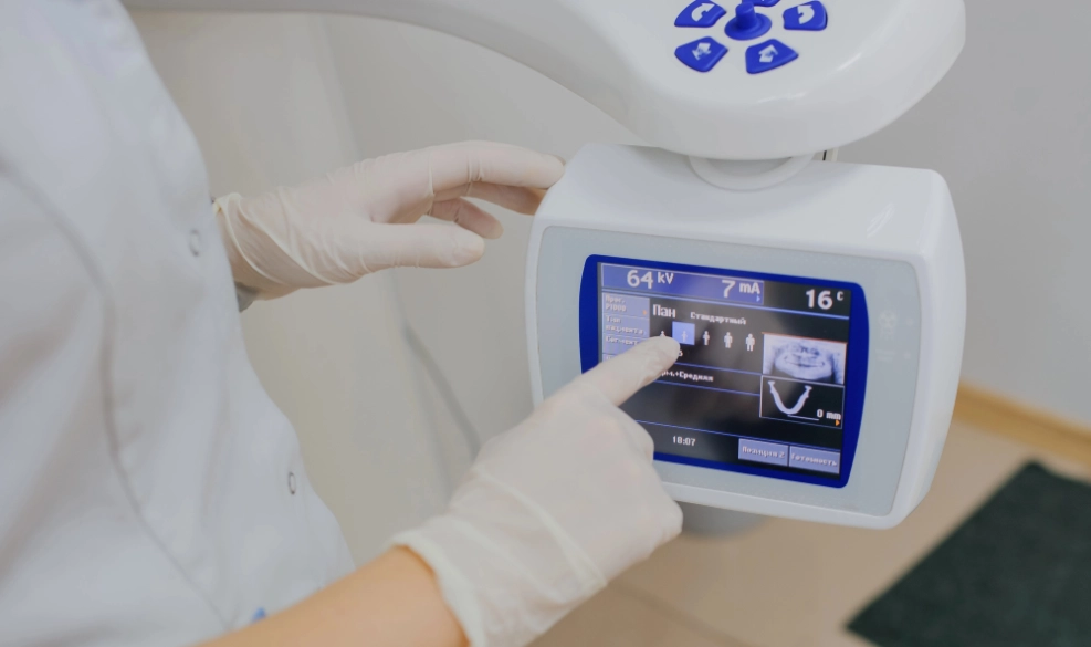
Our CS 9300 cone beam CT system captures highly detailed images while using significantly less radiation than a traditional medical CT scan. Because the scanner can focus on a specific area of concern, we're able to gather the information we need while minimizing unnecessary radiation exposure.

How Does 3D Imaging Work?
During your scan, you'll stand comfortably while the imaging unit rotates once around your head. In just a matter of seconds, the system captures hundreds of individual images that are combined to create a detailed, three-dimensional view of your oral and facial structures.
The process is fast, painless, and non-invasive. Once the scan is complete, Dr. Murray can examine the images from multiple angles, identify concerns that may not be visible on traditional X-rays, and develop a personalized treatment plan based on your unique anatomy.
Because the images are available immediately, we can often explain your diagnosis, discuss treatment options, and answer your questions during the same appointment.


Which Issues Can Be Diagnosed with 3D Imaging?
Three-dimensional imaging allows us to evaluate a wide range of oral and maxillofacial conditions, including:
- Impacted teeth and wisdom teeth
- Bone loss and jawbone defects
- Dental implant planning
- Cysts, tumors, and other oral pathology
- Sinus anatomy and conditions requiring sinus lift procedures
- Jaw injuries and fractures
- Nerve location prior to oral surgery
- Other conditions affecting the teeth, jaw, and surrounding structures
Is 3D Imaging Safe?
Yes. Cone beam CT technology is considered a safe and effective diagnostic tool when used appropriately. Our system is designed to capture detailed images while limiting radiation exposure by scanning only the area needed for your diagnosis or treatment.
Dr. Murray recommends 3D imaging only when the additional information will help improve diagnostic accuracy, treatment planning, or surgical outcomes.


